Research
페이지 정보

본문
Laboratory of Molecular Imaging aNd Therapy Lab, MINT
- Oncology -
02 Research
This laboratory is conducting a research for the treatment of cancer or rare diseases using molecular imaging and nanotechnology to perform translational research to develop diagnostic and therapeutic imaging techniques. using molecular imaging. Our laboratory was established in 1991, and we published about 600 SCI papers to major academic journals, and recognized internationally in the field of molecular imaging research. Through various research projects collaborated with local/foreign researchers, our laboratory has introduced the latest findings to the imaging societies and produced globally talented individuals.
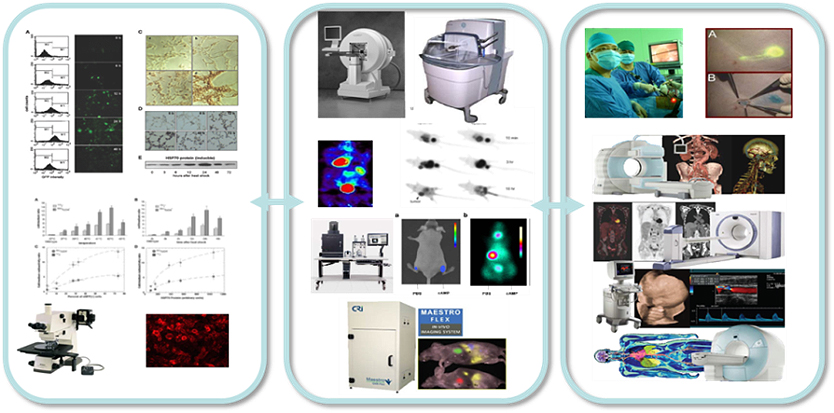
Fig. 1 Various imaging techniques for molecular imaging
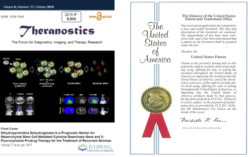
Fig. 2 Cover issues of journals and patent applications
A. Roles of exosomes in the tumor microenvironment
Exosomes are vesicles that are secreted in living cells that contains various types of proteins, miRNA, and signal transmitters; thus, it plays an important role in signal transmission between cells. Exosome has been set as a new paradigm in cancer research by playing a role in tumor microenvironment to tumor proliferation and metastasis. We found that hypoxia-induced exosomes contain miR-210 and involve in angiogenesis (Oncotarget 2016). Our recent researches focus on the roles of radiation-induced exosomes in M1/M2 polarization and EMT/MET transition.
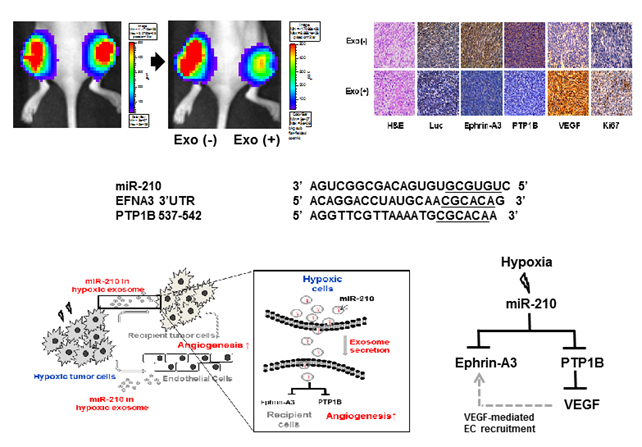
Fig. 3 Hypoxia-induced exosomes transfer miR-210 in tumor microenvironment
B. Enhancing the effect of radiation therapy by increasing the radiation sensitivity
In order to enhance the effect of radiation therapy, we introduced the BRD to the tumor cells. Both in vitro and in vivo experiments, therapeutic efficacy of I-131 was greatly improved in the tumor cells with BRD overexpression compared to the cells without BRD overexpression (Mol Cancer Therapeutics 2015). Our recent studies also focus on the development of new radiation sensitizers.
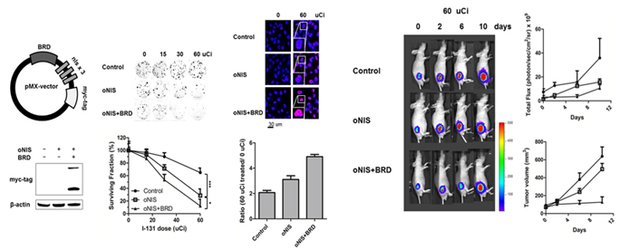
Fig. 4 Evaluation of therapeutic efficacy of I-131 treatment by changing radiation sensitivity
C. Finding new biomarkers for 18F-FDG uptake in tumour cell
18F-FDG PET detects accumulation of an glucose analogue 18F-FDG, and it is highly sensitive and accurate for tumor diagnosis.
Increased 18F-FDG uptake in tumor cells has been reported to be
related to the increased glucose metabolism in tumor cells. Expression of
glucose transporter 1 and hexokinase 2 is important biomarker for positive
signals from 18F-FDG PET, but molecular mechanism of 18F-FDGuptake in tumor cells is not clearly identified. Recently our research focuses
on adenine nucleotide translocase, a protein located at the inner mitochondrial
membrane, and their relationship to 18F-FDG uptake.
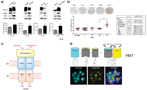
Fig. 5 18F-FDG uptake and ANT expression in different types of cancer cells.
D. Imaging stem cells
We evaluated mesenchymal stem cell (MSC) based glioma therapy using
various imaging modalities. For specific tumor targeting and reducing side
effects, cytosine deaminase (CD) was transduced to MSCs and a prodrug 5-FC was
administrated. In this research we found that our therapeutic MSC/CDs have
ability to chase gliomas and dihydropyimidinde dehydrogenease is a prognostic
marker for MSC/CD therapy (Theranostics 2016). This results were selected as a
cover issue of the journal (2016 Oct).
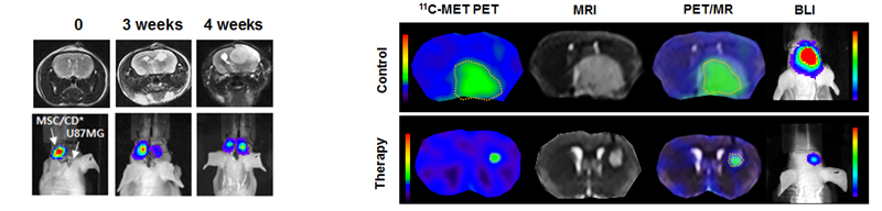
Fig. 6 Evaluation of MSC/CD therapy using molecular imaging
E. Development of optimized reporters
We optimized reporter gene constructs and developed more sensitive reporters, such as luciferases for optical imaging (Mol Cells 2012) and sodium iodide symporters (Theranostics 2015) for nuclear imaging. Our recent projects are focused on the optimization of renilla/gaussia luciferases for optical imaging, transferrin receptors for MR imaging, and generation of reporter-expressing transgenic mice.
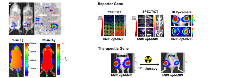
Fig. 7 Development of optimized reporters
F. Imaging Immune Cells
For tracking immune cells, we developed optimized reporter-expressing transgenic mouse and isolated immune cells from the mouse. By adoptive transfer of reporter expressing immune cells, we successfully visualized CD4/CD8 T cell distribution in skin-graft model (Exp Mol Med 2015). Currently, we are applying this reporter mouse to evaluate the efficacy of vaccine candidates.
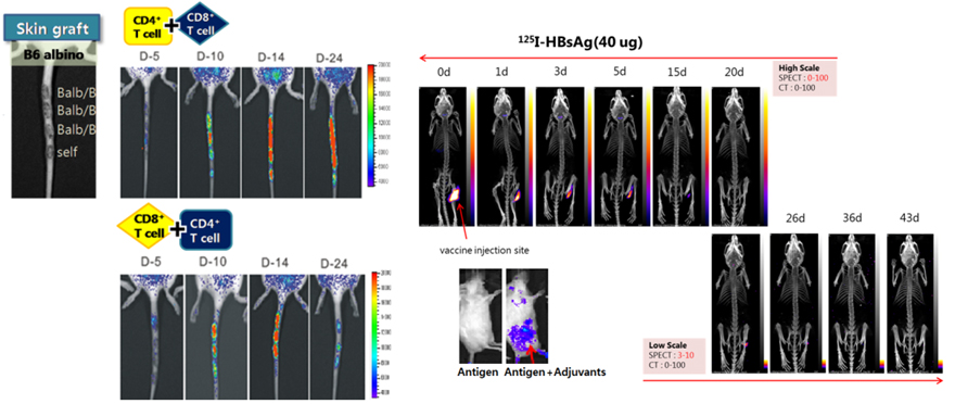
Fig. 8 Immune cell tracking in skin-graft and immunization model
G. Sentinel Lymph Node detection using nanoparticles
For detecting sentinel lymph node detection to remove metastatic tumor, we developed a silica nanoparticles for PET/MR/Optical imaging (Nanomedicine 2012).
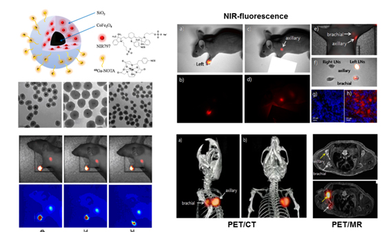
Fig. 9 Sentinel lymph node detection using silica nanoparticles
H. Tumor imaging using gold nanoparticles
For tumor-specific imaging, we developed cRDG-conjugated gold nanoparticles and labeled with radioiodine (Small 2011). Our recent studies focus on the development of encapsulation method for radioiodine on gold nanoparticles.
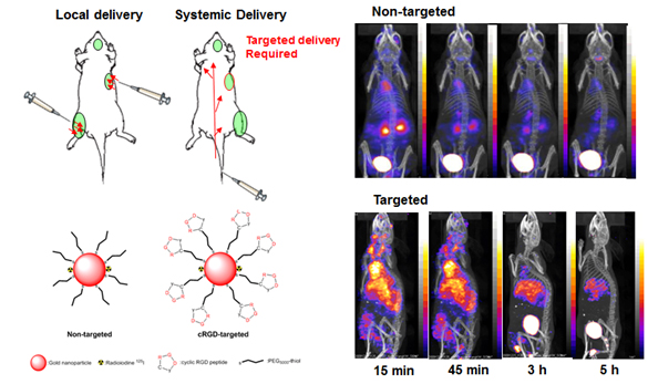
Fig. 10 cRGD-gold nanoparticle to target tumor
I. Tumour-targeting imaging with albumin
For tumor-targeting imaging, we developed cRDG-conjugated albumin to
increase the circulation time in vivo. We successfully visualized tumor
targeting with cRGD-Albumin, and theranostic uses of cRGD-Albumin are under
investigation.
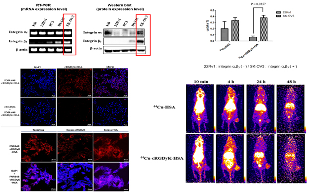
Fig. 11 Tumor targeting imaging with cRGD-albumin
J. Tumor-targeting imaging with Oncolytic vaccinia virus
For evaluating the efficacy of tumor-targeting therapy using oncolytic vaccinia virus expressing cytosine deaminase and somatostatin receptor, optical/nuclear imaging are used for visualizing tumor.
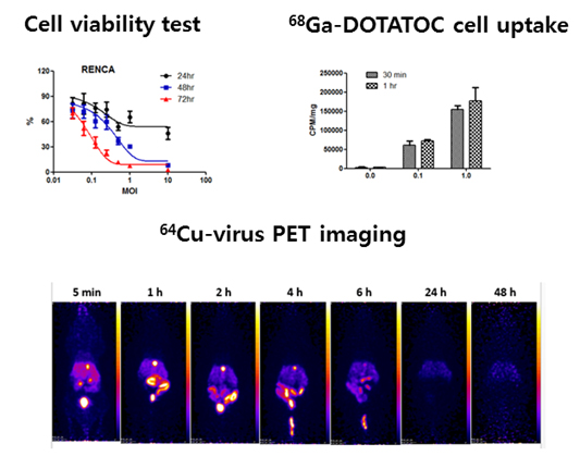
Fig. 12 Tumor targeting imaging with oncolytic vaccinia virus
K. SPARC (Secreted Protein Acidic and Rich in Cysteine) targeting tumor imaging with albumin
We are investigating the distribution of album and SPARC proteins in a mouse xenograft models to evaluate the specificity of albumin in SPARC-expressing tumor.
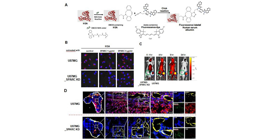
Fig. 13 Binding of fluorescence labeled SPARC and albumin
L. Imaging Rheumatoid Arthritis by tracking inflamed cells
Existing imaging techniques for Rheumatoid Arthritis are limited to visualize structural changes in bones. Our researches adopt new imaging markers to trace inflamed immune cells for visualizing inflamed region.
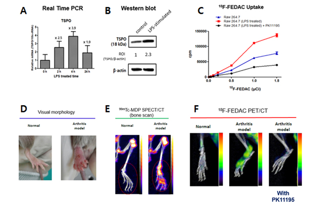
Fig. 14 Imaging inflamed cells with TSPO (Translocator Protein 18kDa) targeting probes
댓글목록
등록된 댓글이 없습니다.